
ELISA with an “S”.
We have all heard a lot about “ELISA” tests, particularly from Dr. Birx over the past several days. I think she uses the term so freely because she has worked in the field for over 30 years and for her the term ELISA falls off the tongue as easily as KLEENEX. For many of us, however the term doesn’t mean much, so I thought it might be useful to explain what the test is and why it is important.
ELISA, is as you probably have guessed, an acronym (isn’t everything these days?). It stands for “Enzyme-Linked ImmunoSorbent Assay”. Don’t worry, it is a lot simpler than it sounds. I will try to explain what it is, why it is so simple and why it will be extremely important in the near future. There is also a closely related test called an RIA (RadioImmune Assay, pronounced “R,I,A”), and you might here that acronym more as the days go by.
The great benefit of these types of tests is that, for the most part they are done in with very small amounts of material, and many tests can be done at one time. These tests are performed in small 96-well plastic plates:
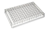
I am pretty sure that you have seen these types of plates in pictures of medical laboratories, usually with a technician filling them with little syringes (those are called pipettes). Sometimes those syringes have multiple outlets so that the technician can fill an entire row at one time.
There are basically 2 ways that tests are done using these plates and as I describe them you will better understand where the names ELISA or RIA come from. For those of you familiar with these tests you will understand that there are multiple variations from what I describe, but I will try to make the descriptions simple.
The basic strategy of tests using these techniques is simple. Something is put in a well that attaches to the sides of the well. Other “reagents” are then added sequentially to the well, and results are then tabulated either by “reading” the wells by eye, or through automated machines. What item is attached to the well, and the order of what is added to it determines what you are testing for.
So, let’s say you want to look to see if there are antibodies to COVID-19 in the blood of a particular individual. Here is how you use immune assays to find out.
First, you put either the virus or pieces of the virus into all of the wells on a plate. It is clearly safer to use pieces of the virus rather than intact virus. This is a good time to talk about what happens when you “kill” a virus particle. As we have talked about in the past, a virus particle (specifically a coronavirus) is a piece of genetic material, in this case RNA, trapped inside a fatty bubble that has a few proteins on its surface. The entire particle, proteins, lipids (fatty envelope) and genetic material needs to be intact in order for the virus to bind to a cell, incorporate into the cell membrane and inject the RNA. If the pieces are broken apart, so that the proteins are separate from the fatty material and the RNA is loose, you cannot have an infection. It is not that an infection is less likely, in fact it is impossible. If you separate the battery and the light bulb from the casings of your flashlight, you can’t have any light, not a chance of having a little light. No light at all is possible.
This is the purpose of washing your hands or using cleaners. Soap dissolves the fatty bubble of the virus, as does 70% alcohol wipes, or bleach.
Getting back under the hood in the lab…. If you place the broken pieces of virus into these plastic plates they will attach to the walls. In most cases the plastic of the wells is sufficient for attachment to take place, in some cases you will need to treat them chemically in advance; however for our discussion let’s assume that when you put the virus pieces, suspended in a liquid, into the wells, a lot of the pieces will attach to the sides and bottom of the wells. You can then rinse out the wells so that nothing remains in the well that is not attached to the walls.
Now you have a set of 96 wells, all of which contain pieces of the virus.
The next thing you do is put the blood (or the serum which is the liquid without the cells from the blood) into the well. You can therefore test 96 different patients in the 96 different wells. (In reality you test fewer than 96 because good science requires duplicate tests, tests with different concentrations of serum called “dilutions”, and a set of control wells in which no blood is added; however, you can still test dozens of individuals per plate). If there are antibodies in the blood that can bind to those virus particles, they will stick on the walls of the wells.
Now the well wall has two layers; the first is the virus pieces, the second is the attached antibodies. For patients without any antibodies that recognize those virus pieces there is only one layer, the virus pieces.
Next, after washing out the blood from the wells, a further reagent is added. This is the one that makes the test. It is another type of antibody, maybe from mice or another animal. The key here is that this antibody recognizes HUMAN ANTIBODIES. It has the ability to bind to human antibodies.
Now the well wall has 3 layers; the virus pieces, then the human antibodies from the blood, then the mouse antibodies that bind to the human antibodies. In the wells where there was no human antibody bound to virus pieces, there is no mouse antibodies attached. This is where the term “immunosorbent” comes from. The immunoglobulin, the antibody is adsorbed to, or in other words attached to the assay plate.
Now for the best part. Before that mouse antibody was added to the plate a special chemical was attached to it. In most cases this is an enzyme. So that when the mouse antibody attaches to the wells with human antibody, it brings with it an enzyme. A good example of this type of enzyme is HRP, horseradish peroxidase. Yes, Virginia, there is a use for horseradish outside of roast beef sandwiches, shrimp cocktails and gefilte fish!
The specific chemistry of how this it is used is not important here. The basic concept is that with this enzyme now attached to the sides of the well, other chemicals can be added that will be broken down by the enzyme. Some of these reactions can result in color changes, some in the creation of solid precipitates that accumulate at the bottom of the well in a visible button, some result in the creation of molecules that are fluorescent. In any of these cases the plate can be placed in a machine and automatically read to give the results from each of the 96 wells.
This allows us to determine which of the blood samples tested had antibodies to the virus pieces.
Sometimes, rather than attach an enzyme like HRP to the mouse antibody, the mouse antibody is “labeled” with a radioactive atom like tritium (radioactive hydrogen). In this case, when the radioactive antibody is attached to the well wall there is no additional chemical reaction necessary, the plate can be put in a special machine that reads the radioactivity of each well. Only those wells in which the radioactive mouse antibody was attached will be read as positive.
When an assay uses an enzyme like HRP, it is doing and “enzyme-linked” assay. Hence the name ELISA; when the assay uses a radio-labelled antibody, it is called a “radio immune” assay, hence RIA.
BUT WHAT IF YOU WANT TO LOOK FOR VIRUSES RATHER THAN ANTIBODIES?
You can use the same type of assay, but the layers are different.
In this case you take antibodies to the virus itself and attach them to the walls of the well. The best reagents here are “monoclonal antibodies”, and maybe in a subsequent post I will explain what they are (I did most of my research creating, studying and using monoclonal antibodies).
With those antibodies attached to the well walls you can now add the blood from a patient. If there is virus present it will attach to the antibodies on the wall creating a second layer.
Now you add another batch of antibodies (these could be human, or from another animal, it doesn’t matter as long as they can bind to the virus. The virus is now bound on one side to the walls through antibodies, and the other side is bound to the new antibodies. These new antibodies can have the same types of chemicals attached to them as the mouse antibodies described above. The plates can be “read” in the same way as above. The only difference here is that you are looking to see if there is virus in the blood rather than antibodies in the blood.
These assays can be done in a few hours, and there is very little limit to how many you can do at once. There are plates that have even more wells in them, and machines that can read plates quickly.
Although these tests are not designed for point of care use, and won’t be used at your drive thru, they have extraordinary power to provide studies of large populations.
I hope this helped you understand the terms that we are hearing more and more.